Treatment of pregnancy-associated breast cancer
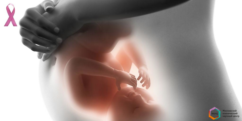
NewsPregnancy – associated breast cancer (BC) is a malignant tumor detected during pregnancy or within one year after delivery. It accounts for 25% of all malignant diseases during pregnancy and is more common than others.
The vast majority of cases (70-90%) are represented by invasive flow tumors. The neoplasm is initially localized in the milk ducts, then spreads to the surrounding tissues and affects the lymph nodes.
Symptoms
The clinical picture of breast cancer in pregnant women does not differ from the manifestations of the tumor in non-pregnant women. In the early stages, there are no symptoms. As the tumor grows there are changes in the mammary glands that are noticeable to the woman:
- painful tightness in the chest or armpit;
- changing the form;
- discomfort in the nipple area, discharge.
Diagnostics
The doctor who discovered signs of breast cancer during the reception of a pregnant patient, sends her for examination. Diagnostics include:
- Ultrasound of the mammary glands, regional lymph nodes;
- mammography (in the second and third trimester of pregnancy) with screening of the fetus;
- magnetic resonance imaging (without contrast during pregnancy, with contrast after delivery);
- Core-a biopsy for histological verification of the diagnosis.
Among women, there are biases about the dangers of X-ray examination of the mammary glands during pregnancy. They often lead to the refusal of mammography. But, according to the American Association of Obstetrics and Gynecology (ACOG), a radiation load of less than 0.05 Gy does not pose a danger to the fetus. During pregnancy, there is a natural change in the structure of the mammary glands. Therefore, 3D mammography (tomosynthesis) should be preferred. The radiation load of this study does not exceed 0.003 Gy.
Another reliable method of studying the neoplasm is core-biopsy. It is used for the collection of tumor material and further study. The method allows you to accurately make the correct diagnosis and prescribe a course of adequate therapy. The procedure is low-traumatic and safe for both the patient and the fetus.
Treatment
The patient management process includes:
- Discussion of the decision to maintain or terminate the pregnancy by doctors, the patient and her family.
- Decision to terminate the pregnancy-treatment begins immediately and in full after the abortion.
- The decision to save the pregnancy-treatment is carried out by permitted methods.
The difficulty of treating breast cancer during pregnancy is to achieve an optimal therapeutic effect with minimal negative impact on the fetus and the outcome of pregnancy. Treatment tactics are selected individually in each specific case. The attending physician takes into account the prevalence of the tumor and the duration of pregnancy.
Treatment is carried out in the following sequence:
Stage 1-radical mastectomy. The operation and the anaesthetic aid are relatively safe for the mother and child. Greater harm to the fetus is caused by the mother's stress, hypoxia and hypotension during surgery. Therefore, the doctor should keep a clear control over the patient's condition during the operation and eliminate negative conditions in a timely manner.
Specialists do not support performing organ-preserving surgery in the first and second trimester of pregnancy, as subsequent radiation therapy of the remaining part of the breast is indicated.
Stage 2-radiation therapy and chemotherapy.
In pregnant women, radiation therapy is not used because of its teratogenic properties. The threshold damaging dose for the fetus in the first and second trimesters is 0.1 Gy. It leads to the following negative consequences:
- spontaneous miscarriage;
- birth defects;
- delayed neurocognitive development;
- leukemia.
Today, specialists can calculate the risk of ionizing effects on the fetus taking into account the indicators of the fetus, the methodology and dose of radiation, the size of the breast. The radiation field can be more accurately focused on the breast, chest, and lymph nodes. Also, the use of protective equipment for the uterus will help to further reduce the dose of radiation exposure by 50-75 %. This calculation model is used in clinical situations that require a decision to maintain pregnancy against the background of treatment of breast cancer diagnosed in the first or second trimester of pregnancy.
The third trimester is characterized by a lower sensitivity of the fetus to radiation exposure. However, today it is allowed to plan a radical operation with radiation of the breast after delivery, if the malignant neoplasm was detected in the third trimester.
The course of chemotherapy can be carried out no earlier than the second trimester of pregnancy, when the fetus is formed and the main organs and systems have been laid. The risk of developing birth defects during chemotherapy in the first trimester reaches 20%.
The accumulated experience of using chemotherapy for breast cancer from the second trimester of pregnancy demonstrates its safety during intrauterine development and in the neonatal period. However, in order to avoid the development of hematological complications (neutropenia, thrombocytopenia, anemia) during childbirth and in the early postpartum period, a second course of chemotherapy should not be carried out after 35 weeks of pregnancy or within 3 weeks before the planned delivery.
The chemotherapist will select the right drug therapy with drugs that do not have negative effects on the fetus and the duration of pregnancy.
Special attention should be paid to pregnancy planning for women who carry mutations in the BRCA 1/2 genes, since there is a high probability of developing breast cancer during pregnancy. We recommend that you undergo a comprehensive examination and consultation with a mammologist before pregnancy.